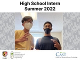
CAST sets high bar for High School senior's lab internship
Colin Wang,CAST Intern from Marriotts Ridge High School
Colin Wang, senior at Marriotts Ridge High School in Marriottsville, MD completed an internship in summer 2022 at the Center for Advanced Sensor Technology (CAST) at Chemical Biochemical and Environmental Engineering Department at UMBC. Wang was mentored by Christopher Slaughter, '23, M31 computer engineering and supervised by Dr. Govind Rao, professor in the Department of Chemical, Biochemical and Environmental Engineering.
The main purpose of Wang's summer internship was to gain research experience with real research goals and to work on automatic biosensors for glucose monitoring. The biosensor detects and reads the concentration of glucose in a liquid sample without the need of diabetic patients to constantly prick their skin. Colin Wang's internship immersed him in a new research topic while also teaching him what being in a real lab was like and giving him a sense of family.
Colin shared his experience the write up he titled "CAST & Curious - How a Summer Internship in Biotechnology Shaped Me". Learn more about other opportunities for High School students at CBEE : https://cbee.umbc.edu/highschools-visits/
CAST & Curious - How a Summer Internship in Biotechnology Shaped Me
by Colin Wang
Part 1: Introduction
Nothing beats in-person, real experience. High school classes and labs, while great at propelling a student into biotechnology, are only a stepping stone towards real lab experience with real research goals. My internship in UMBC's Rao Lab with the members of CAST immersed me in a new research topic while also teaching me what being in a real lab was like and giving me a sense of family. Thus, the present paper aims to summarize the research I participated in at CAST as well as to review perhaps the best summer experience possible for an ambitious student.
Part 2: Automatic Glucose Biosensor
- Context:
My main connection to CAST came through the Automatic Biochemistry Analyzer, also known as the Automatic Biosensor. Simply put, when given a sample, a matching protein, and the correct column preparation, the biosensor detects and reads the concentration of a molecule, such as glucose, in a liquid sample. Why is this needed? Current methods of glucose monitoring in human blood require diabetic patients to constantly prick their skin, which is uncomfortable and painful. Alternatively, noninvasive glucose monitoring by passive diffusion, or without breaking the skin, would give convenience and ease to the 37.3 million2 diabetic patients in the US. This aim is best achieved through transdermal patches that, when placed on the skin, collect enough sweat to then be analyzed by the automatic biosensor.
- Physical Description:
What is the biosensor? It's a system, a compact box, that consists of a syringe, four valves, tubing, a fluorometer, a microdialysis device, and beakers for waste and Phosphate Buffered Saline (PBS). (CAST has two versions, one black and one white. The black system, an earlier prototype, is slightly outdated but is often preferred for its functionality. The white system also uses a different column shape.)
- Backbone:
The backbone of the biosensor is that a protein reacts with the sample molecule and, with the help of a light, produces a fluorescent reaction, which is then quantified and displayed graphically on a computer screen. For the glucose biosensor, the logic follows: more glucose, more reaction, more fluorescence. Therefore, greater values of fluorescence intensity indicate higher concentrations of glucose.
- Experiment Preparation:
The protein (Glucose Binding Protein (GBP)) is labeled with a Fluorophore, or a polarity-based dye (BADAN), at a specific Cysteine mutation in the protein code so that it will react with the glucose. Nickel-NTA (Ni-NTA) beads work in tandem with the protein to produce the reaction. (In the experiments I ran, we tested for ATP rather than glucose, so we used a different binding protein.) Both the protein and Ni-NTA beads are loaded into a microcolumn, a plastic rectangle-shaped device with an "IN" fitting and "OUT" fitting. The microcolumn, or column, serves as the protein's and beads' home as well as a chamber for the PBS/sample to flow through. (See Column Packing for ATP Biosensor, CAST protocol.) A "Column Wash" on the "Standalone Functions" page is run to flush out old, leftover protein. Finally, the container of PBS 1X stock is refilled. (To make new PBS, mix 90mL diH2O from the sink nozzle in the "Glassroom" with 10mL PBS from Chad's room. Use the labeler to mark the date that the PBS was renewed.)
- Experiment Running:
Experiments3 run by either withdrawing or infusing PBS stock into/from the main syringe. Withdrawing PBS from the stock container refills the syringe, if it is empty. Infusing PBS, on the other hand, pushes liquid through the biosensor's tubing system of four valves. Using the "Manual Control" page to flick valves by hand is always available, but using the controls on the "Standalone Functions" page saves time. The system directs the sequence automatically.
- Valve Functions:
- Valve 1 connects the syringe to either the PBS container or the system tubing.
- Valve 2 directs the PBS to either the microdialysis device (to pick up sample) or the microcolumn (to flush out leftover protein).
- Valves 3 and 4 direct PBS with sample to either waste or the column (to react with protein).
The tubing sequence for an experiment, assuming the column has been cleaned and prepared, is as follows: PBS is infused from the syringe to the microdialysis device, where it picks up glucose from a sweat sample, then runs through the microcolumn, reacts with the protein, and travels to waste. A blue/violet4 light is shined at the column, the resulting fluorescence is picked up by the fluorometer, and the results are displayed graphically on the computer screen.
- Steps:
- Refill - PBS is withdrawn from container.
- Sample - PBS is infused to sample, collects glucose through microdialysis device pores.
- Measurement - Mixture moves to column, reacts to protein and light, produces fluorescence.
- Purge - Old liquid is flushed out of tubing.
- Graph Interpretation:
Data comes in the form of spikes (maximums) and valleys (minimums). The baseline value refers to the average bottom value, or the average resting value of fluorescence intensity. Each peak value shows the highest concentration of glucose for that round. The "Last Peak Amplitude" value is a measurement of how much above the baseline the last measured peak was.
- Troubleshooting:
Carrying out experiments isn't all sunshine and roses, though. Things can be confusing. For example, it took a few puzzling minutes and a quick conversation with Hasib, Chris' mentor, for us to learn that the frits pushed into the "IN" fitting of the column (4.4mm) were actually of a smaller diameter than the normal filters built into the column channel (5.6mm). (Building more frits is easy—just use the hole puncher in the "Glassroom" to cut down the 5.6mm ones.) Mistakes are also commonplace, but expected. We once inserted the frit before the protein, making us have to break open the frit with tweezers. Another time, I accidentally let the Ni-NTA beads settle before pipetting them into the column, clogging the "IN" fitting and forcing us to have to break open the column with an Exacto knife.5
Larger problems sometimes require a bit more thinking. When the biosensor wasn't detecting enough ATP in our E. coli/Lysogeny Broth sample, we tried pipetting BugBuster solution into the sample, thinking it would lyse the cell walls and release more cellular ATP, which the microdialysis device would then pick up. When the peaks were still low, we added more. Several unsuccessful rounds later, we tried something different: we cleansed the microdialysis device itself with a round of plain PBS. Our peaks shot up! It had turned out the BugBuster had indeed lysed the E. coli cells, but the broken cell organs had then clogged the microdialysis device and blocked ATP from going through. A rush of PBS had unclogged it. Troubleshooting and finding innovative solutions like those was frustrating but worth the trouble.
Part 3: Being in Lab
Arguably just as important as the research content itself was the experience of being in a real lab.
- Patterns of Lab:
To begin, start with the patterns of lab CAST taught me, or how things run in a professional lab setting. Over time, for example, I became familiar with each room's purpose. New columns are laser cut in a room dubbed the "Workshop," beakers are washed and autoclaved in the "Glassroom," and no one's allowed in the room labeled "Vida's Room!"6 I also learned the more random things, like the chart that assigns people to wash glassware on different days, or where the Ni-NTA beads are stored (the top shelf of the main room's mini fridge), or that the lights have to be dimmed when dealing with proteins to prevent denaturation (even if it's hard to see). There are days for experiments, and then there are days for writing (Chris much prefers the former). And apparently, the Rao Lab in summer is much less frantic than during the semester, when deadlines are harder and the pressure is greater.
- Family:
More unexpected was the familial comfort I grew to have around everyone I met. All the people I met were so nice, and simply seeing them on a daily basis and learning different things from each person really infused a sense of family into me. That was the sense I got from CAST. They were one family, and they welcomed me with open arms.
It was Trish greeting me as I walked in, Chris chuckling about "the kids these days" (he denies being Gen Z one moment, then fanboys over Tony Stark the next), Chad laughing with me about the sorry state of the proteins fridge (he writes his name on the boxes he uses, but a good 50% of the contents are frozen over by ice), Shayan casually doing a gel electrophoresis while talking with me about his PhD, Hasib helping Chris with the LabU code, and Dr. Rao popping into our office to say hi. It was being trusted to run experiments myself and meeting the new kid Elias (who's still a year older than me). It was Chris and I on some days getting lunch from The Hub (he insisted on paying, citing "mentor's duty") and then on some days looking up at 2pm during experiments and realizing we forgot to eat lunch again. Even at its most basic level, it was driving to lab each day, knowing that I had my own desk and crew waiting there to say hi. The sense of family I felt at CAST gave me another perspective on what a career in research had to offer, beyond the research itself.
Part 4: Conclusion
In more ways than this report can put into words, my experience at CAST was invaluable. Learning about a research topic and carrying out experiments showed me the value of doing meaningful, if hard, work. Being forced to troubleshoot when problems arose gave me first-hand examples of grit and creative thinking. Watching and picking up on the customs of a real lab put me in the closed-toed shoes of a researcher. And finally, being welcomed into the tight-knit community of CAST gave me an appreciation for colleagues as friends.
CAST, you set the bar incredibly high for every other lab experience in my future. I hope to bring the lessons you taught me wherever I go. Special thank you to Dr. Hennigan and Dr. Rao for giving me this amazing opportunity, and to Chris for being there with me every step of the way. Thanks guys, and cheers.
<><><><><>
Posted: February 24, 2023, 2:46 PM
